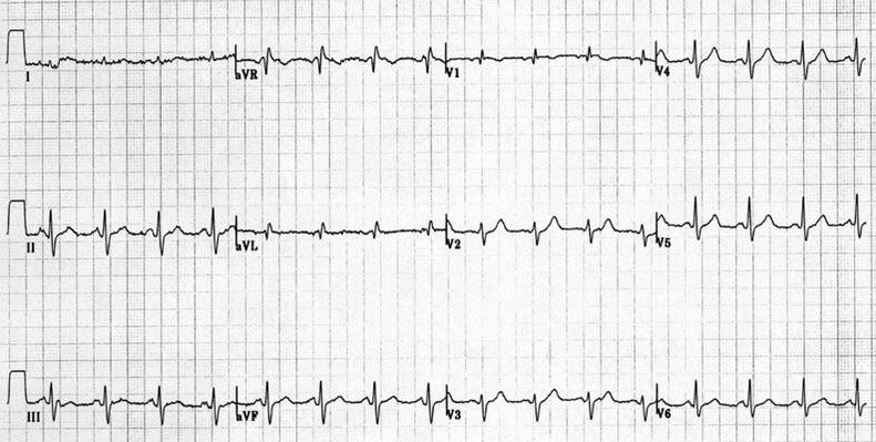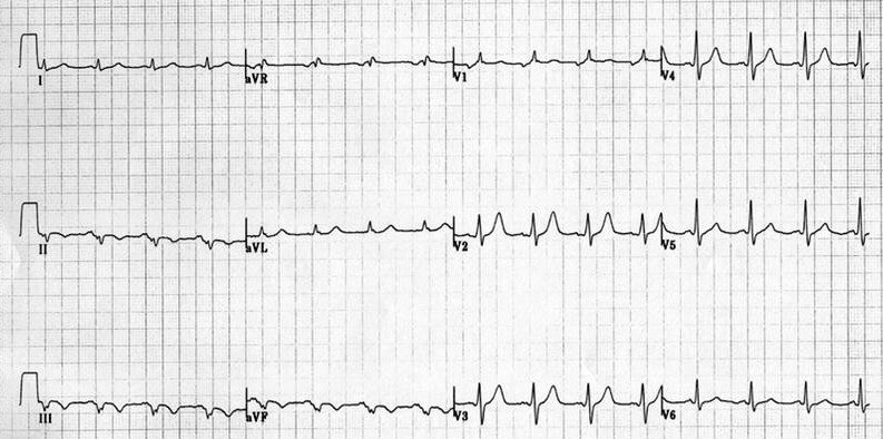
sent me the following ecg's and said at the time "I was amazed many of my patients could be cured of their inferior wall MI by just moving the lower limb leads from the limbs to the torso."
The "standard" configuration used was with the lower limb leads at the ankle and upper limb leads on the clavicle at the mid-clavicular line, with the patient supine.
(It must be noted that since these were to be serial ECGs, they would be done with the patient walking, jogging or even running on the treadmill. Therefore, to minimize muscle artifact all electrodes were placed over bone. All precordial leads were shifted upwards from their usual intercostal space placement to a rib.)
The "modified" configuration was as above, but with lower limb leads attached on the torso, mid- clavicular, at the bottom of the rib cage and with the patient standing on the treadmill just prior to starting their walk. What with their age (and therefore, elastic skin) I could not guarantee where the electrodes ended up after changing from the supine to standing positions.